To advance a personal injury or medical practice claim, it’s crucial to convey the key medical details of the case in a comprehensible but sensitive manner. Medical animations have emerged as a powerful communication tool to support litigation efforts. Using spoken words alone is a gamble in the courtroom, where participants can fall behind – not understanding, nor relating to, the technical concepts being discussed. This can lead to disengagement and compromised objectivity. Medical animation companies can minimize this risk with clear demonstratives that keep a non-technical audience attentive and following along.
Visually engaging solutions keep the jurors in the know, helping to clarify in-depth expert testimony. Artery Studios, a renowned medical animation company, has revolutionized trial presentations and gained recognition by offering accurate scientific depictions that promote informed and fair litigation outcomes.
Best Practices for Creating Effective Animations for Trial
To integrate legal animations seamlessly into case strategy, medical animation companiesmust ensure the presentations align with litigation strategies. They consider numerous factors, including:
Scientific accuracy: To withstand scrutiny from opposing counsel, medical animations must be backed by scientific facts from not only the file, but also the opinion of medical experts and respected literature. A reputable medical animation company will appreciate an attorney’s approach and collaborate with consulting experts to ensure the legitimacy of all details portrayed.
Visual clarity: Unnecessarily complex visuals can cloud perception, deterring people from understanding the critical issues being litigated. Medical animations help pinpoint key considerations through focused design. Color, contrast, close-up views, camera pans, labels and clear anatomical portrayals integrated into each movie, provide communication clarity to maximize understanding.
Courtroom admissibility: Animations must adhere to legal standards to be admissible as demonstrative evidence. It’s essential that medical animation companies maintain a non-prejudicial approach while assisting the attorney in presenting their position – be it plaintiff or defense. Medical animators ensure these visuals are court-admissible by strict adherence to relevancy, dovetailing exactingly with the documented case record and opinion of the experts, and vetting the demonstratives by the testifying experts who will verify their accuracy and utility.
Customization: Every case comes with a unique set of facts and contested elements. Legal animations are tailored to portray the specific injury or medical condition being litigated on that file. Through customization, a medical video animation company helps strengthen the case by presenting a direct ‘translation’ of the case record.
Engaging storytelling: A compelling narrative is critical to drive juror engagement. Following a logical sequence of visual narrative,medical animations articulate the mechanism of injury, surgical issues, medical malpractice factors, or other case elements stepping through the issues to maximize understanding by the jury.
By adhering to these practices, medical animation companies produce demonstratives that are not only visually appealing but also strategically designed to optimize trial outcomes.
Communication Considerations for Jury Understanding
The essence of legal animations is their ability to engage jurors and make complex proceedings accessible to those with limited knowledge of the subject matter. Medical video animation companies create visuals that resonate with jurors, by taking several factors into account:
Pacing: What a medical expert may take hours to explain, a carefully crafted medical animation can make clear in minutes or even seconds. An appropriately paced animation with periodic pauses for emphasis, allowing for the expert to provide further explanation, is a requisite to help a non-technical audience absorb information comfortably.
Scene complexity: Medical animations are structured to demonstrate one concept at a time, with camera moves, zooms, titles and labels appearing sequentially (rather than simultaneously). These movies avoid cluttered or overly technical portrayals, being streamlined to avoid cognitive overload for the viewer. In this way, the most relevant details of the case are conveyed without delving into unrelated or superfluous matters.
Emotional resonance: Too often, scientific subject matter can be unrelatable to someone with limited training or understanding. Visuals that depict complex topics in a simplified but realistic style are more relatable, allowing the opportunity for the jury to connect with the issue at hand. A medical video animation company translates potentially triggering gruesomeness that viewers may want to turn away from, into sensitive portrayals that they welcome engaging with.
Cultural sensitivity: Jurors come from varying backgrounds, with distinct life experience, education and perceptions. Medical animation services make use of universally relatable visual cues and design principles to safeguard against heuristic discounting of the animated content, optimizing the educational value of these demonstratives.
Integration of Animations with Expert Testimony
The persuasiveness of medical animations in personal injury and malpractice proceedings is elevated further when integrated into expert witness testimony. Not only is this essential for admissibility of the demonstratives, but it allows for a cohesive approach, allowing the evidence to unfold seamlessly. Considerations for this integration include:
Reinforcing expert narratives: Animations serve as essential communication support to reinforce an expert’s testimony, making abstract concepts readily understandable. For example, a surgeon testifying about an incorrectly performed procedure can utilize a medical legal animation to more clearly communicate the intricate details of the anatomy, surgical approach, and actions taken that caused the outcome.
Enhancing less than clear testimony: When faced with an expert who lacks charisma, is a poor communicator, or who engages in complex medicalese, a well-crafted medical animation can optimize their communication style, allowing them to be a much better teacher/educator.
Interactive storytelling: Well-crafted medical animations allow the testifying expert to add commentary to the visual in real-time. Scene titles that separate concepts, breaking sequences into stages and incorporating pauses in the footage, enable them to add a live ‘voice-over’, enriching understanding by the judge and jury.
Pre-testimony collaboration and validation: A reputablemedical video animation company works in cooperation with case experts to verify all content, facilitating their testimony. Credibility of the demonstrative and its admissibility is maximized by this collaboration, along with the expert’s signoff of the visuals created.
Engagement of the jury over lengthy expert testimony: The extensive explanation of complex medical concepts can challenge the attention span of jurors. This is of particular concern with younger jurors (so-called ‘digital natives’) accustomed to multitasking and quickly swiping screens. Expertly constructed animations strike a balance of presenting complex information quickly enough to engage the viewer, but slowly enough for all levels of education and lived experience to appreciate. While reducing cognitive overload, visuals presented in a motion format, with an evolving timeline, retain their attention.
Optimizing Settlements
Medical animations should be integrated into case development early – including at the deposition and mediation stage. The following case study showcases the power of a medical video animation company to optimize early outcomes:
Case Study: Dental Surgery Leading to Nerve Injury
Context: Artery Studios was retained by the plaintiff’s lawyer on a case involving an improperly performed dental surgery that resulted in significant damage to a nerve supplying the teeth and adjacent area. When installing a crown, the oral surgeon failed to properly insert a dental implant into the jawbone, resulting in pain, numbness, tingling, burning and itching in the plaintiff’s skin and lips. The plaintiff’s lawyer required a demonstrative to show how drilling to create a bony socket for the implant was done improperly, causing damage to the inferior alveolar nerve. The implant selected by the doctor, was also argued to be too long, causing further compromise of the canal in which the nerve ran. Intricate anatomical and surgical concepts needed to be clearly explained for the jury to fully appreciate all relevant details.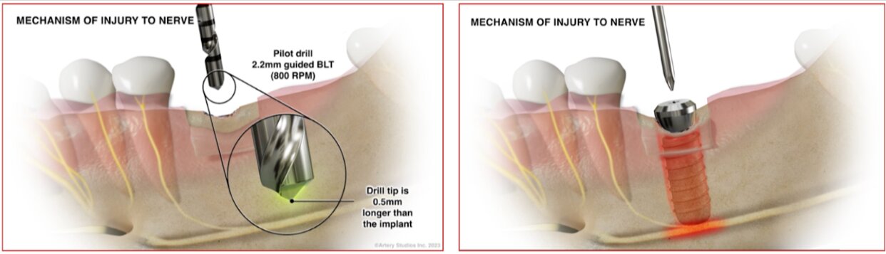
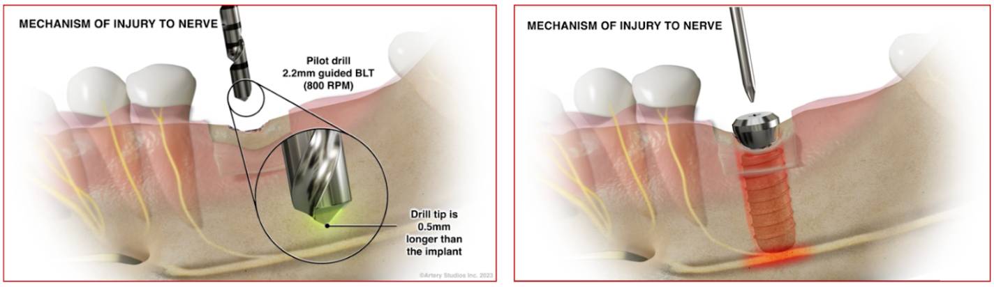
Figure 1: Frames from an animation showing details of the drill bits used to create a hole in the jawbone for a titanium post, concepts of implant insertion and associated nerve injury.
Artery Studios’ contribution: Stephen Mader and his team at Artery Studios created a customized medical legal animation explaining the anatomical, surgical and complication issues. Animated segments demonstrated the course of the inferior alveolar nerve in the mandible (lower jawbone), showing what areas it innervates, the configuration of the drill bits used to create a socket in the bone for the tooth replacement, and the details of the oversized implant inserted (Figure 1).
The chosen form of demonstrative for this case was animation due to the contentious motion-related factors that needed to be explained. Integration of camera motion and zooms, as well as portrayal of action of the dental devices and insert placement, would help engage the jury and assist them in grasping the dynamic surgical process that caused this irreversible harm.
Outcome: The animation helped clarify critical details, facilitating settlement before the case proceeded to trial. The persuasive and concise video helped all litigation participants fully appreciate critical issues related to this surgery. The law firm was very pleased with the settlement outcome, praising Artery Studios on the visual strategy and execution.
Have a Complex Case?
Medical video animation companies like Artery Studios have shown, time and again, that accurate, substantiated, client-specific animations that integrate seamlessly with expert testimony and present medical evidence in an impactful and clear visual manner, can maximize litigation outcomes.
As legal animationsadvance with changing technology,they will continue to support courtroom narratives with efficient communications strategies. Partnering with a trusted medical animation company like Artery Studios can make a critical difference in the outcome of complex cases.


